牛津仪器集团成员
扩展

Innovative 3D Neuron Tracing with Imaris

The Imaris for Neuroscientists package is the perfect combination of tools for researchers working in various neuroscience disciplines. Imaris enriches advanced 3D/4D visualization and analysis methods with Filament Tracer - best in class software for an automated detection of neurons including spines and other filamentous structures in 2D/3D or 4D datasets, measurements, object tracking, plotting, group comparisons with statistical tests and two-way interface for customization in Matlab, Java or Python.

Request Pricing

Tracing neurons and detection of cells and structures

Microscopy image analysis requires precise detection of multiple biological structures based on the initial signal saved as an image data. Object detection in Imaris will be quick and easy as users are guided within a wizard driven interface to create perfect models of their data. With our patented Torch™ tool and several performance improvements tracing neurons with Autopath or Autodepth in dense and thick samples is very efficient and becomes a unique experience.

Automatic Tracing

Automatic neuron tracing is guided within the wizard based interface – starting point and seed points along the filament are detected with just few mouse clicks.

 
  • Dedicated algorithms available for trees without loops or structures which may contain loops.
  • Adjust multiple parameters to ensure the best possible detection quality: amend automatically calculated threshold, add/delete start or end points of the filament.
  • Segmenting images even with sub-optimal quality (e.g. overall low signal to noise ratio and/or in the presence of high intensity (non-filament) points in the background).
  • All dendritic spines along the filament and creates a new class of objects

Semi-automatic Autopath

Autopath method is recommended for tracing filamentous structures in big datasets of multiple GBs in size (up to 1 TB tested) and dense networks like cleared samples, cleared tissues, entire brains or vasculatures.

 
  • User only selects the start and end points in 3D while the best fitting filament path is calculated and highlighted.
  • Tracing can be paused after every intermediate step and started again from the selected end or branching point.
  • The filament diameter is automatically calculated from the structure
  • Autopath method is equipped with tools which enable tracing in within dense neural networks: Torch, Depth Visibility.

Semi-automatic Autodepth

Another method recommended for analysis of big datasets of multiple GBs in size (up to 1 TB tested) and dense networks like cleared samples, cleared tissues, entire brains or vasculatures.

 
  • User draws a line along the structure in the 3D image which is automatically centred within the nearest filament structure (highest data intensity).
  • Drawing can be done in multiple small steps to ensure the best possible fitting
  • Filament diameter is automatically calculated from the structure.
  • Autopath method is equipped with tools which enable tracing in within dense neural networks: Torch, Depth Visibility.

Object Detection Spots and Surfaces

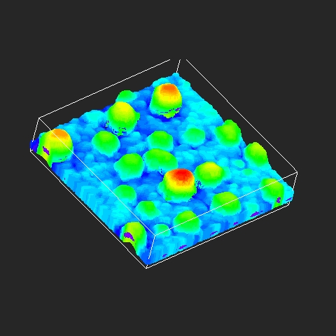
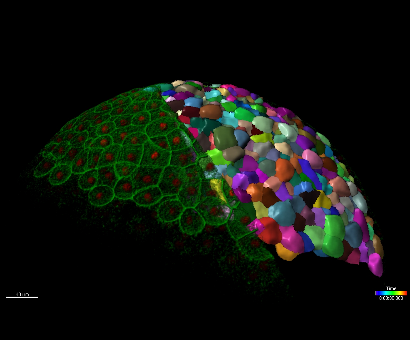
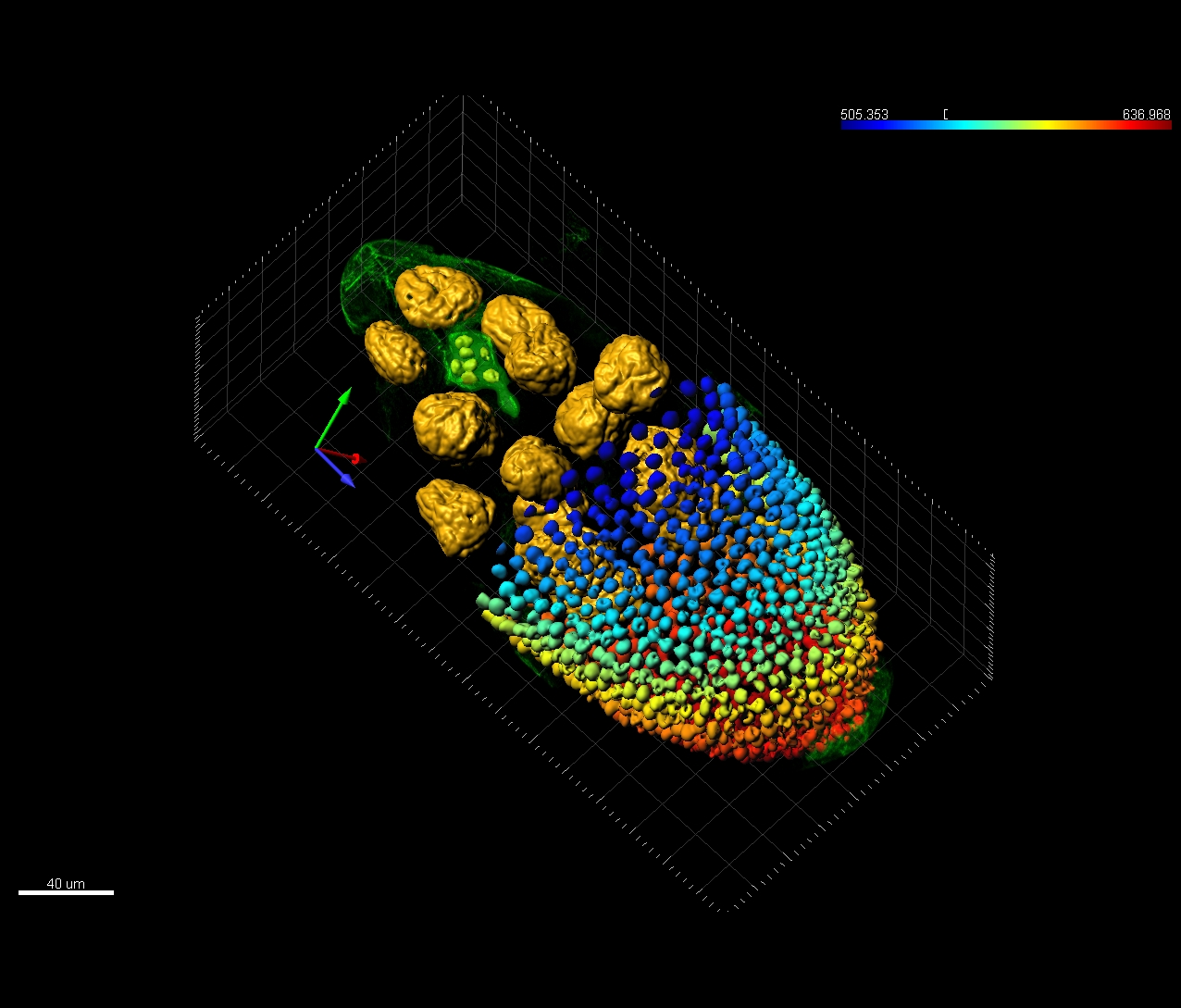
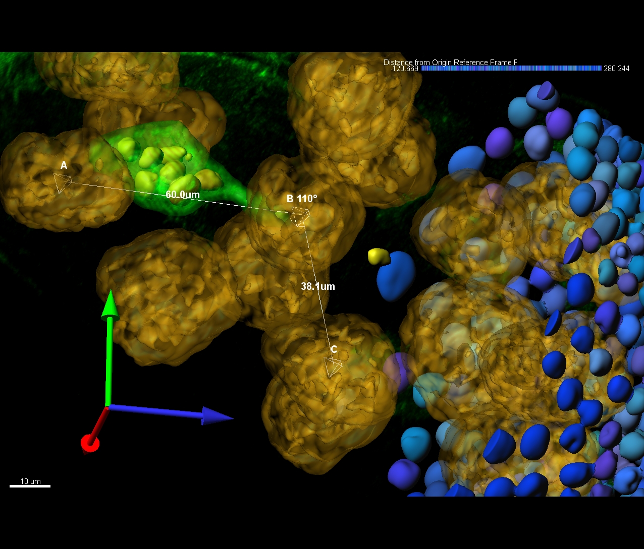
Surface objects can be created over the boundaries of objects of different shapes and sizes, like cells, nuclei, nucleoli, brain structure or a filament while the Spots objects are used to model point-like or vesicle-like structures in the data and to quickly identify thousands of structures.

 
  • Surface and Spots objects creation is guided within the wizard based interface
  • Surfaces and Spots can be edited, added or deleted in 3D space
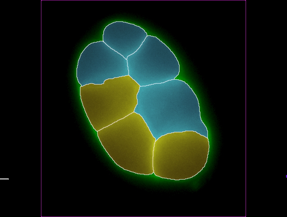
  • Surfaces can be drawn on 2D planes as a contour iso-line, magic wand or free-hand and recreated as 3D objects
  • Several statistics like number of objects, position, volume, area, elipticity, fluorescence intensity inside the structures, and many more are automatically calculated
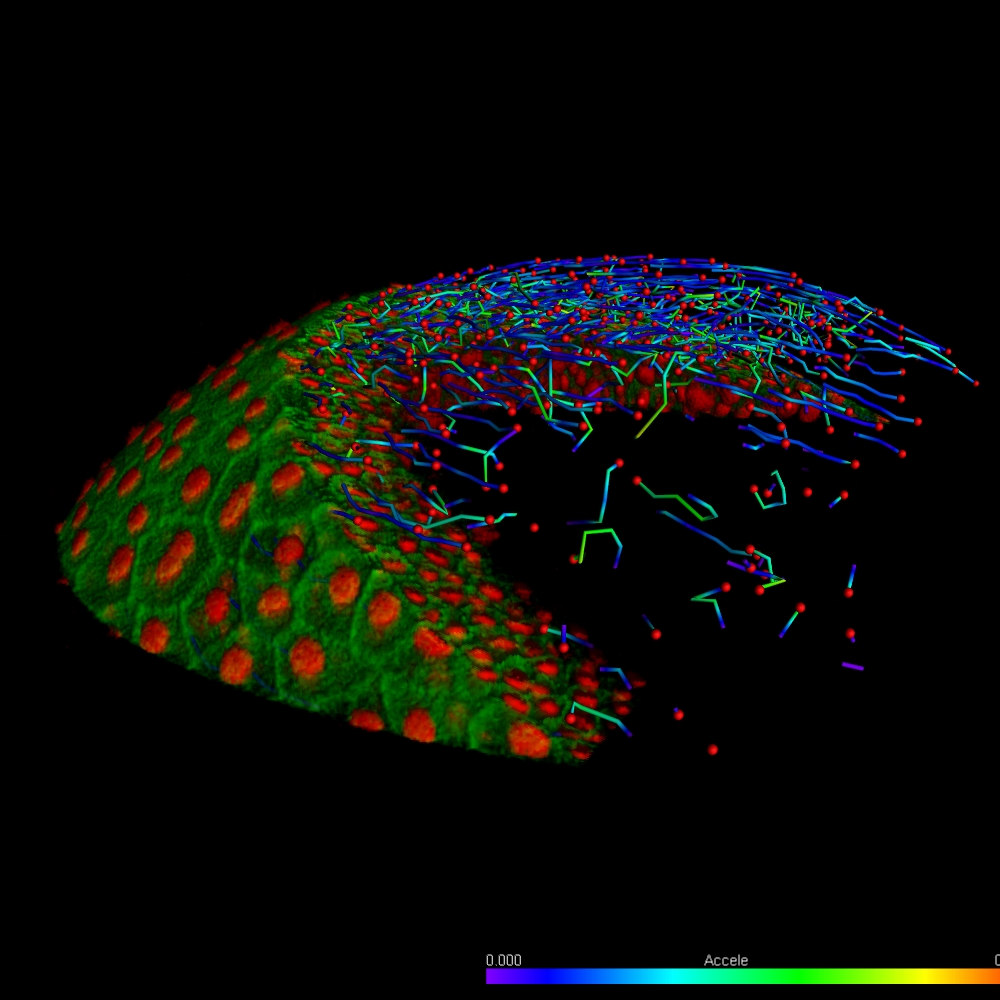
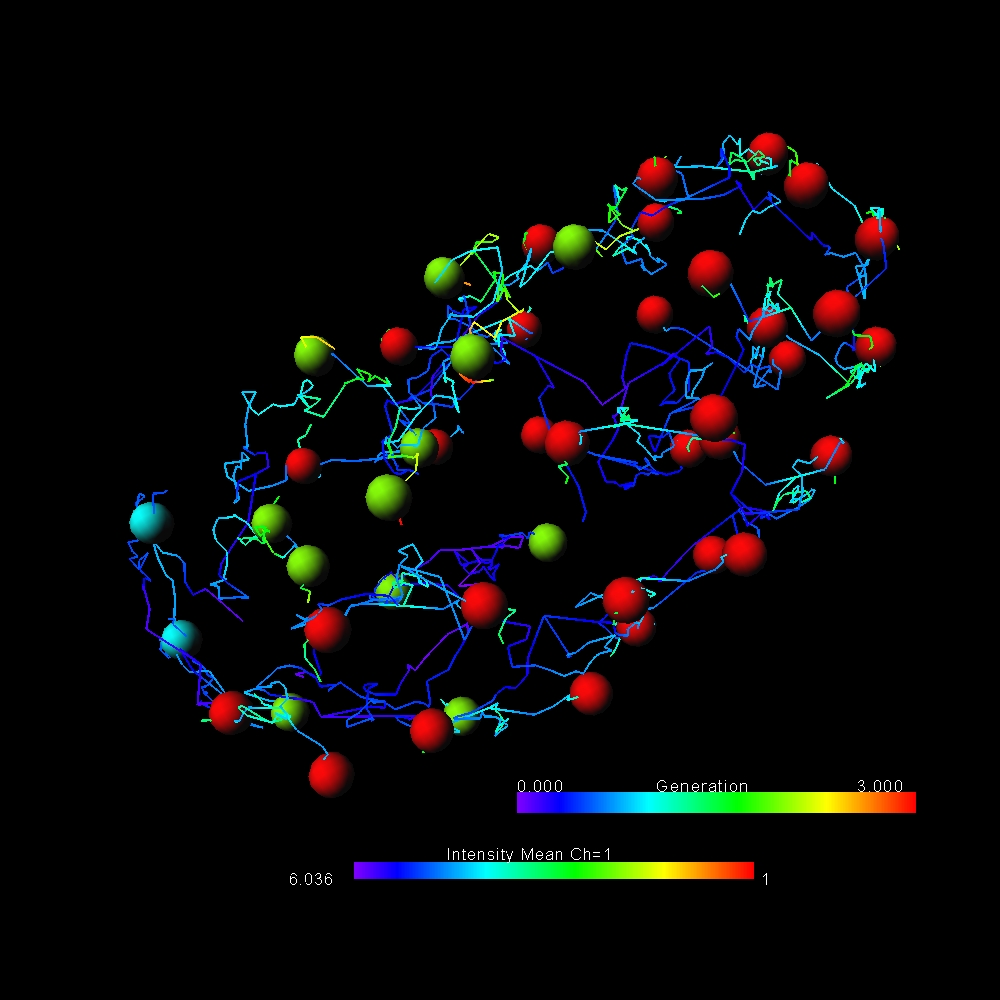
  • Surface and Spots Objects can be tracked in time series which allows monitoring of the temporal changes in shape or intensity and gives several motion related statistics like speed, acceleration, displacement, track path, lineage (for dividing objects)

Dendritic spine detection and classification

Regardless of which filament tracing method is chosen (wizard driven automatic tracing or an Autopath/Autodepth mode) dendritic spines can be automatically detected and precisely modelled based on the threshold.

 
  • Full set of statistics including spine number per segment, length, volume, area, diameter
  • Dendritic spines can be classified based on their morphological features like length, head and neck diameter (ie. mushroom, stubby, filopodia)

Visualization and animation export

Imaris provides a complete set of features for visualization of multi-channel microscopy datasets from static 2D images to 3D time series regardless their size and format. Using Imaris for Neuroscientists users can:

  • Organize and manage full experiments including images and data analysis in Imaris Arena.
  • Use premier 3D/4D volume rendering modes: (MIP, Blend Projection, Shadow Rendering, Normal Shading)
  • Benefit from Imaris high performance IMS file format which guarantees smooth navigation even in very large 3D datasets (TB range).
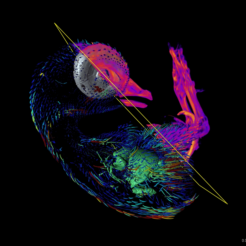
  • Combine volume rendering, object representation, clipping planes and cross-section slices.
  • Make high quality snapshots ready to be used in publications in Snapshot Tool.
  • Easily make animations which include volume rendering, detected objects, annotations, object motion by adding several frames of interest in a Key Frame Animation Tool

Statistics and measurements

After segmenting the image data Imaris calculates a wide range of statistics for all detected objects: Filaments, Surfaces and Spots. All values can be used for color coding, plotted inside Imaris (using Vantage plots) or exported in an .csv or .xls file format. Parameters presented below are the most common statistic types needed by biologists. Imaris reports many more.

Motion Analysis
  • Length
  • Mean Diameter
  • Branching Angle
  • Spine Density
  • Resistance
Cells
  • Dendrite Branch Points
  • Dendrite Terminal Points
  • Sholl Intersections
  • Spine Terminal Points
  • Dendrite Branch Level
Spots
  • Length
  • Volume
  • Mean Diameter
  • Area
  • Terminal Point Diameter
Surfaces
  • Area
  • Volume
  • Intensity
  • Position (x, y, z)
  • Number (counting objects)

Further benefits of Imaris for Neuroscientists

This package is about much more than just tracing. With the inclusion of various Imaris modules, you can experience complete analysis freedom during your experiments.

Plotting and Data Comparison

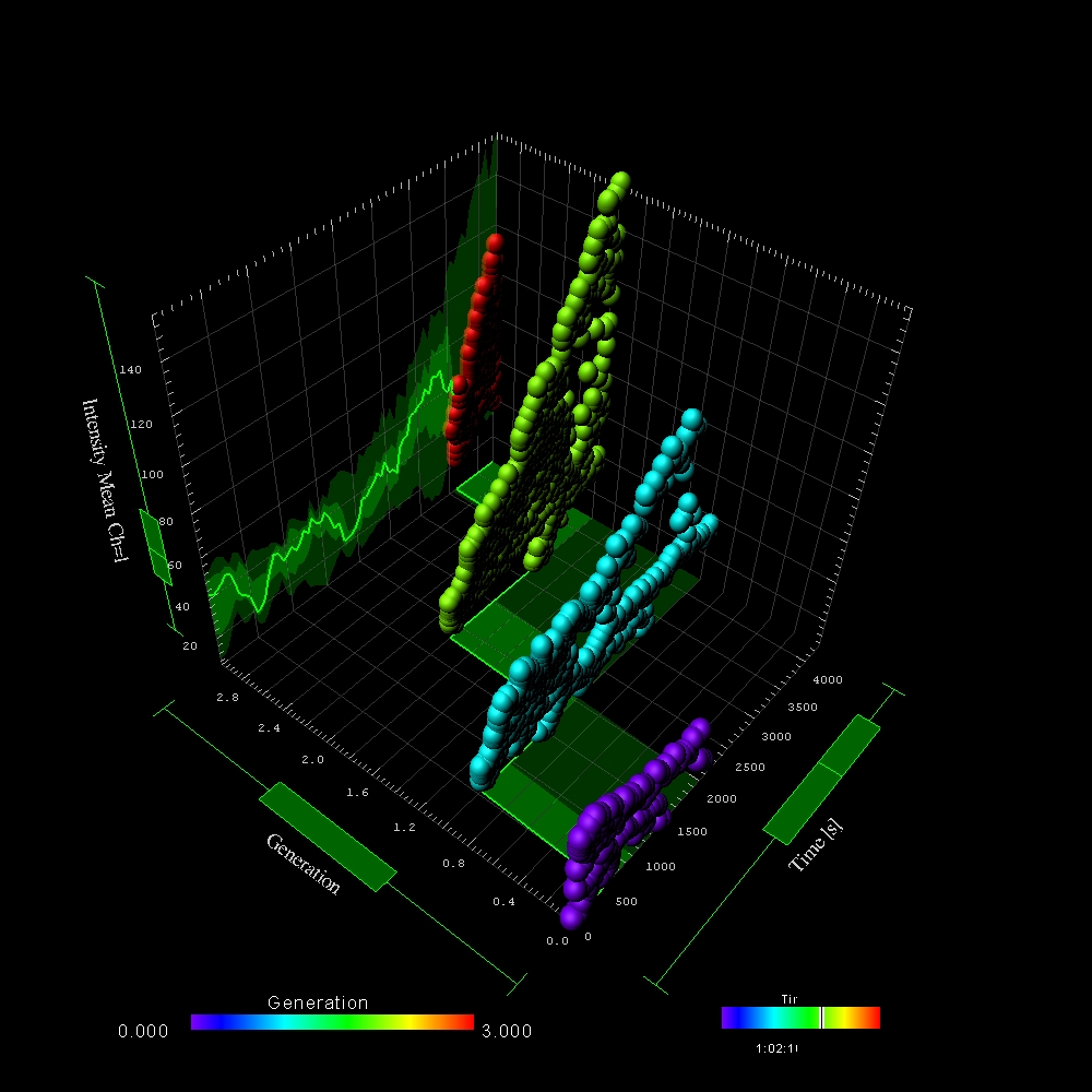
Imaris Vantage plotting tool enables to visualize segmented objects on multi-variate scatterplots to discover hidden relationships between object statistics.

  • Up to 5 variates represented on xyz axes, size and color) or create a gallery of all detected objects (like spines or dendrites) to compare their shapes.
  • Two way interaction between original image and the plot: when the group of objects selected on the plot, it’s also highlighted on the original image.
  • Easily compare chosen statistics between your control and experimental groups on uni- or multi-variate scatterplots.
  • Test statistical hypotheses using: t-Test F-test, Kolmogorov-Smirnov, Wilcoxon.
  • All created plots are ready to use in papers and presentations - you can export them with Snapshot.
 

Tracking Algorithms

Imaris Track included in Imaris for Neuroscientists offers a variety automatic tracking methods which can best fit the type of motion.

  • Five sophisticated tracking algorithms which can also handle objects that appear or disappear between frames.
  • Uses previous object speed and directionality for predicting future positions as well as weighted intensity information to enable the most accurate tracking possible
  • Intuitive and flexible track correction tool for editing individual tracks once the automatic tracking is finished
 

Two Way Interface for Customization

Imaris XT module is particularly useful for scientists who write image analysis application code in Matlab or Python and for everybody who has sophisticated image analysis needs which are not fully covered by Imaris main functionality. Imaris XT provides:

  • Access to the pre-existing library of XTensions – custom image analysis scripts which can be modified for specific applications.
  • Two-way interface from Imaris to classic programming languages: Matlab, Java or Python and an image export/import to Fiji.
  • Rapid development and integration of custom algorithms which exceed the possibilities of generic image processing and are tailored to very specific scientific applications.
open-source-coding

Additional Resources

The Imaris Learning Center hosts a wide range of tutorial videos, how-to articles and webinars to guide you through the many features of Imaris. We have provided some links below which will get you started on some of our most recent developments.
沪ICP备17031777号-1 公安机关备案号31010402003473